 ЖИ көмекші


Diseases of the nervous system ant their treatment презентация

 Жүктеу
#1 слайд
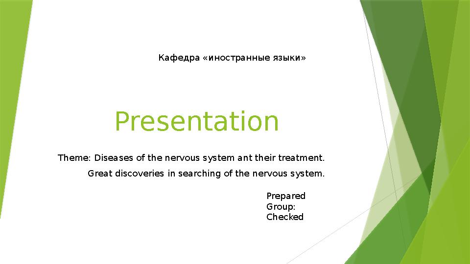
Presentation
Theme: Diseases of the nervous system ant their treatment.
Great discoveries in searching of the nervous system. Кафедра «иностранные языки»
Prepared
Group:
Checked
1 слайд
Presentation Theme: Diseases of the nervous system ant their treatment. Great discoveries in searching of the nervous system. Кафедра «иностранные языки» Prepared Group: Checked

#2 слайд
Plan:
I.Introduction
II.Main part
• Sympathetic Nervous System
• Parasympathetic Nervous System
• Peripheral Nervous
III.Conclusion
IV.Bibiliography
2 слайд
Plan: I.Introduction II.Main part • Sympathetic Nervous System • Parasympathetic Nervous System • Peripheral Nervous III.Conclusion IV.Bibiliography

#3 слайд
Introduction
The nervous system includes the brain, spinal cord, and a
complex network of nerves. This system sends messages back
and forth between the brain and the body. The brain is what
controls all the body's functions. The spinal cord runs from the
brain down through the back. It contains threadlike nerves that
branch out to every organ and body part. This network of
nerves relays messages back and forth from the brain to
different parts of the body.
3 слайд
Introduction The nervous system includes the brain, spinal cord, and a complex network of nerves. This system sends messages back and forth between the brain and the body. The brain is what controls all the body's functions. The spinal cord runs from the brain down through the back. It contains threadlike nerves that branch out to every organ and body part. This network of nerves relays messages back and forth from the brain to different parts of the body.

#4 слайд
Main part
Billions of neurons work together to create a communication network.
Different neurons have different jobs. For example, sensory neurons
send information from the eyes, ears, nose, tongue, and skin to the
brain. Motor neurons carry messages away from the brain to the rest
of the body to allow muscles to move. These connections make up the
way we think, learn, move, and feel. They control how our bodies
work — regulating breathing , digestion , and the beating of our hearts
4 слайд
Main part Billions of neurons work together to create a communication network. Different neurons have different jobs. For example, sensory neurons send information from the eyes, ears, nose, tongue, and skin to the brain. Motor neurons carry messages away from the brain to the rest of the body to allow muscles to move. These connections make up the way we think, learn, move, and feel. They control how our bodies work — regulating breathing , digestion , and the beating of our hearts

#5 слайд
What Are the Parts of the Nervous
System?
The nervous system is made up of the central nervous system and the peripheral
nervous system:
The central nervous system includes the brain and spinal cord.
The peripheral nervous system includes the nerves that run throughout the whole
body.
The nervous system uses tiny cells called neurons (NEW-ronz) to send messages
back and forth from the brain, through the spinal cord, to the nerves throughout the
body.
5 слайд
What Are the Parts of the Nervous System? The nervous system is made up of the central nervous system and the peripheral nervous system: The central nervous system includes the brain and spinal cord. The peripheral nervous system includes the nerves that run throughout the whole body. The nervous system uses tiny cells called neurons (NEW-ronz) to send messages back and forth from the brain, through the spinal cord, to the nerves throughout the body.

#6 слайд
Sympathetic nervous system
Sympathetic neurons have cell bodies located in the intermediolateral columns, or
lateral horns, of the spinal cord. The presynaptic fibers exit the spinal cord through
anterior roots and enter the anterior rami of T1-L2 spinal nerves and onto the
sympathetic trunks via white rami communicantes. From here, the fibers may ascend or
descend the sympathetic trunk to a superior or inferior paravertebral ganglion,
respectively, pass to adjacent anterior spinal nerve rami via gray rami communicantes,
or cross through the trunk without synapsing and continue through an abdominopelvic
splanchnic nerve to reach prevertebral ganglia. Because of the central location of the
sympathetic ganglia, presynaptic fibers tend to be shorter than their postsynaptic
counterparts.
6 слайд
Sympathetic nervous system Sympathetic neurons have cell bodies located in the intermediolateral columns, or lateral horns, of the spinal cord. The presynaptic fibers exit the spinal cord through anterior roots and enter the anterior rami of T1-L2 spinal nerves and onto the sympathetic trunks via white rami communicantes. From here, the fibers may ascend or descend the sympathetic trunk to a superior or inferior paravertebral ganglion, respectively, pass to adjacent anterior spinal nerve rami via gray rami communicantes, or cross through the trunk without synapsing and continue through an abdominopelvic splanchnic nerve to reach prevertebral ganglia. Because of the central location of the sympathetic ganglia, presynaptic fibers tend to be shorter than their postsynaptic counterparts.

#7 слайд
Parasympathetic nervous system
Due to the expansive nature of the vagus nerve, it has been described as an ideal “early
warning system” for foreign invaders as well as for monitoring the body’s recovery. Up
to 80% of vagal fibers are sensory and innervate nearly all major organs.
Parasympathetic ganglia have been found to express receptors for interleukin-1, a key
cytokine in the inflammatory immune response.This, in turn, activates the
hypothalamic-pituitary-adrenal axis and SNS, leading to the release of glucocorticoids
and NE, respectively.Studies have correlated inhibited vagal action through vagotomy
and cholinergic inhibitors with significantly reduced, if not eliminated, allergic,
asthmatic, and inflammatory responses.
7 слайд
Parasympathetic nervous system Due to the expansive nature of the vagus nerve, it has been described as an ideal “early warning system” for foreign invaders as well as for monitoring the body’s recovery. Up to 80% of vagal fibers are sensory and innervate nearly all major organs. Parasympathetic ganglia have been found to express receptors for interleukin-1, a key cytokine in the inflammatory immune response.This, in turn, activates the hypothalamic-pituitary-adrenal axis and SNS, leading to the release of glucocorticoids and NE, respectively.Studies have correlated inhibited vagal action through vagotomy and cholinergic inhibitors with significantly reduced, if not eliminated, allergic, asthmatic, and inflammatory responses.

#8 слайд
Peripheral nervous system
The peripheral nervous system derives from neural crest cells. The neural
crest is divided axially into the cranial, vagal, truncal, and lumbosacral
neural crest cells. Truncal neural crest cells contribute to the dorsal root of
the spinal cord and the sympathetic ganglia. The parasympathetic
innervation of the heart forms from the vagal neural crest.The majority of
the parasympathetic nervous system, including all of the ganglia of the head,
has been shown to arise from glial cells, rather than neural crest cells
8 слайд
Peripheral nervous system The peripheral nervous system derives from neural crest cells. The neural crest is divided axially into the cranial, vagal, truncal, and lumbosacral neural crest cells. Truncal neural crest cells contribute to the dorsal root of the spinal cord and the sympathetic ganglia. The parasympathetic innervation of the heart forms from the vagal neural crest.The majority of the parasympathetic nervous system, including all of the ganglia of the head, has been shown to arise from glial cells, rather than neural crest cells

#9 слайд
Conclusion
Dorsal nucleus: provides parasympathetic output to the viscera
Nucleus ambiguus: produces motor fibers and preganglionic neurons that innervate the
heart
Nucleus solitarius: receives afferents of taste sensation and that from viscera, and lastly
Spinal trigeminal nucleus: receives information of touch, pain, and temperature of the
outer ear, the mucosa of the larynx, and part of the dura
9 слайд
Conclusion Dorsal nucleus: provides parasympathetic output to the viscera Nucleus ambiguus: produces motor fibers and preganglionic neurons that innervate the heart Nucleus solitarius: receives afferents of taste sensation and that from viscera, and lastly Spinal trigeminal nucleus: receives information of touch, pain, and temperature of the outer ear, the mucosa of the larynx, and part of the dura
 | Nemours KidsHealth Clevelan")
#10 слайд
Bibiliography
https:// www.ncbi.nlm.nih.gov /books/NBK539845/
Nervous System (for Parents) | Nemours KidsHealth
Cleveland Clinichttps :// my.clevelandclinic.org
› 21...Nervous System: What It Is, Parts, Function & Disorders
Wikipediahttps ://en.m.wikipedia.org › wikiNervous system
National Institute of Child Health and Human Development (.gov)https:// www.nichd.nih.gov › neuroWhat
are the parts of the nervous system?
10 слайд
Bibiliography https:// www.ncbi.nlm.nih.gov /books/NBK539845/ Nervous System (for Parents) | Nemours KidsHealth Cleveland Clinichttps :// my.clevelandclinic.org › 21...Nervous System: What It Is, Parts, Function & Disorders Wikipediahttps ://en.m.wikipedia.org › wikiNervous system National Institute of Child Health and Human Development (.gov)https:// www.nichd.nih.gov › neuroWhat are the parts of the nervous system?


 Жүктеу
шағым қалдыра аласыз


 Бұл курс Қазақстан Республикасы Оқу-ағарту министрлігімен келісілген


