ЖИ көмекші


 Қауіпсіз төлем

жүктеу мүмкіндігіне ие боласыз
Бұл материал сайт қолданушысы жариялаған. Материалдың ішінде жазылған барлық ақпаратқа жауапкершілікті жариялаған қолданушы жауап береді. Ұстаз тілегі тек ақпаратты таратуға қолдау көрсетеді. Егер материал сіздің авторлық құқығыңызды бұзған болса немесе басқа да себептермен сайттан өшіру керек деп ойласаңыз осында жазыңыз
 12 Наурыз 2019
 418

1 жыл бойы шексіз жүктеңіз
Electrical processes in the heart that includes electrocardiography
|
||||||
11.2B Transport of substances
|
School: |
|||||
Date: |
Names of teachers:
|
|||||
Grade: 11 |
Number present: |
Absent: |
||||
Theme of the lesson |
Electrical processes in the heart that includes electrocardiography |
|||||
Learning objectives that are achieved at this lesson(Subject Programme reference) |
Learners should be able to:
|
|||||
Lesson Objectives |
Learners will be able to:
|
|||||
Success Criteria |
The student should be able to:
|
|||||
Language objectives |
Learners can: explain orally, in written formthe electrical pathway of the heart and components of the electrocardiogram Vocabulary: Electrocardiography / ECG, P,QRS,T – waves; nerve impulses to/from the heart; ion movement within myofibrils involved in myogenic contraction, non-conducting layer between atria and ventricles; spread of electrical activity around the heart, waves of depolarisation and time delays device, electrodes, filters, normal ECG, electrical axis of heart, electrical activity of the heart, the potential difference, electrocardiogram, conductive gel, EEG machine, connecting wiresrecords, equipment, amplifiers, output device, wrist, ankle, bipolar leads, lightheadedness, consciousness, data acquisition computer Planned usage Electrocardiography is … By electrocardiography … can be seen … … of the … is represented bythe wave/complex/segment/interval.
|
|||||
Values instilled at the lesson |
|
|||||
Cross-curricular links |
The working of the ECG is based on the principles of Physics (electricity). |
|||||
ICT skills |
The power-point presentation will help students improve their ICT skills. |
|||||
Previous learning
|
The following learning objective has been covered in the previous lesson.
|
|||||
Courseofthelesson |
||||||
Planned stages of the lesson |
Planned activities at the lesson |
Resources |
||||
Start 0-10mins
|
G Activity1 Irashnee
Learners are to be separated into 3 groups of 2 each and 1 group of 3.
Group 1:
INSTRUCTIONS:
Group 2
Group 3
Group 4 Zhanargul Use the Kobra 4 apparatus to record ECG of your partners and record your results.In this work it is necessary to take into account that the ECG data are not used in medicine for diagnosis but only for training
REPORT BACK AND DISCUSSION Irashnee
Group one will be informed that the human heart actually is able to pump 5 liters of blood through the heart in a minute.
Group two will explain how difficult it was to continue squeezing the ball for a minute. This activity would have given them an idea as to how hard the heart actually works in our bodies.
Group 3 will explain the number of counts of their members. This will lead to a discussion on what a heartbeat actually is and what a heartbeat indicates about ones health levels.
Group 4 will report back their blood pressure readings and we can ask learners to hold on to that result till the end of lesson one
|
|
||||
Middle 3 mins
10-12 mins
3 mins
8-10 mins |
Activity 2. Irashnee and Zhanargul A large outline of the heart is put up on the table. Learners are given labels and asked to come up and label the various parts of the heart ( Right and Left atrium , Right and left ventricles, Septum , Sino atrial valve, Superior and Inferior Vena Cavae, Pulmonary artery and Pulmonary Veins and Aorta, Bicuspid and Tricuspid Valves ) Students sign the structure of the heart on paper A3 and then the teacher provides them with a ready drawing of the structure of the human heart so that they can test themselves and adjust their answers and records Activity 3. Zhanargul
view video " work of the heart"
heart modeling in groups /students use foam rubber, fabric, threads, markers, scissors, drawing the structure of the heart/to show the principle of the heart in stages While students make a model of electrical conductivity, the heartbeat teacher tests their work and listens to students ' explanations Sequence of steps in the electrical process of the heart
Activity 4. Irashnee https://en.wikipedia.org/wiki/File:ECG_principle_slow.gif View the animation of the heart and how to write an ECG.After watching the animation, the teacher asks questions. QUESTIONS TO BE ASKED
Activity 5. Irashnee and Zhanargul
A small formative assessment where students can carry out self-evaluation by answering key questions MARKING MEMORANDUM
b)QRS- VENTRICULAR SYSTOLE c) T- VENTRICULAR DIASTOLE d) Q- PRE-VENTRICULAR CONTRACTION e) R-PEAK OF VENTRICULAR CONTRACTION f) S- REPOLARISATION OF VENTRICLES 1.2 AFTER SA NODE CONTRACTS 1.3 – MEASURES RHYTHM OF HEARTBEAT - CAN INDICATE SIZE AND POSITION OF CHAMBERS - DAMAGE TO MUSCLES - EFFECT OF DRUGS - FUNCTIONING OF PACEMAKER 2.1 ELECTRODES 2.2 EACH SHOWS A DIFFERENT REGION OF THE HEART 2.3 CAN INDICATE EXACT AREA THAT IS DAMAGED 2.4 5=60 4,25= x 51
|
https://en.wikipedia.org/wiki/File:ECG_principle_slow.gif
|
||||
Finish 3 mins |
Reflecting IReflective sandwich. |
template of sandwich, stickers |
||||
Extra information |
||||||
Differentiation – how do you plan to give more support? How do you plan
to challenge the more able learners? |
Cross-curricular links Health and safety regulations ICT skills Values instilled at the lesson |
|||||
Differentiation in activities during the lesson. Use of support for students with different perception channels (visual, audial, kinesthetic) Support for weaker students: Translated key words.Individual attention. Advanced students had extra tasks in the worksheet |
Health and safety: work with laptops, movement in the classroom Values links The students will understand the importance and relevance of ECG in their daily lives. Other subject links: The working of the ECG is based on the principles of Physics.
|
|||||
Assessment – how are you planning to check students’ learning? |
Observation, questioning, success criteria for group activity, providing models, worksheet. |
|||||
Reflection
Were the lesson objectives/learning objectives realistic? Did all learners achieve the LO? If not, why? Did my planned differentiation work well? Did I stick to timings? What changes did I make from my plan and why? |
Use the space below to reflect on your lesson. Answer the most relevant questions from the box on the left about your lesson. |
|||||
Learning and lesson objectives were realistic? 90% of learners achieve the LO up to the end of the lessons (they finished the worksheet tasks)
Differentiation worked well. It linked types of activities, perception channels, students’ level in biology. Yes, I stuck to timings I didn’t make changes at this lesson, but for the next time I’ll give the texts to weaker students during listening task. |
||||||
Summary evaluation What two things went really well (consider both teaching and learning)? 1:Activity Whiteboard Writing Race 2:Activity “That Squigglely Line - What Does It Really Mean?” What two things would have improved the lesson (consider both teaching and learning)? 1: To give the texts about ECG to weaker students. 2: To add “2 stars and 1 wish” for asses the models ECG form. What have I learned from this lesson about the class orachievements/difficulties of individuals that will inform my next lesson? To give the texts about ECG to weaker students due to their difficulties in listening video. |
||||||
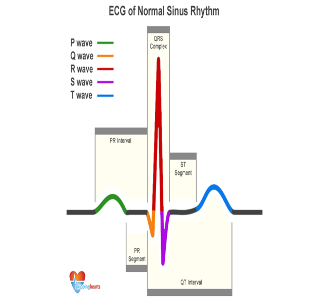 he
P wave represents the spread of a stimulus through the atria
(atrial depolarization).
ACTIVITY : ELECTROCARDIOGRAM
AN ELECTROCARDIOGRAM IS USED TO RECORD THE ELECTRICAL ACTIVITY OF THE HEART. MAKE A CAREFUL STUDY OF A PORTION OF AN ECG AND ANSWER THE QUESTIONS THAT FOLLOW:
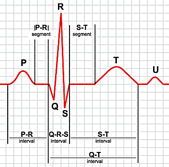
Which important processes in the heart is being depicted by:
P wave ___________________________
QRS interval ______________________
T wave ___________________________
Q ____________________________
R ____________________________
S ____________________________
At which point does a P wave begin ? ________________________________________________________________________________________________________________________________
Name the cycle that is covered by the ECG . ________________________
Name the 3 phases involved in the above cycle.
_________________________________________
_________________________________________
_________________________________________
Briefly describe what is happening during each of the above phases.
________________________________________________________________________________________________________________________________________________________________________________________________________________________________________________________________________________________________________________________________________________________________________________________________
State functions of :
SA- NODE: ____________________________________________________________________________________________________________________________
AV NODE :
____________________________________________________________________________________________________________________________
Pukinjje Fibres:
____________________________________________________________________________________________________________________________
State any FOUR uses of an ECG.
________________________________________________________________________________________________________________________________________________________________________________________________________________________________________________________________________________________________________________________________
This is an actual ECG
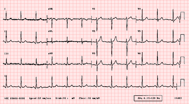
What are the electrical leads used during an ECG called ?
________________________________________________
What do the different V values stand for ? ___________________________
Why are they so important in an ECG ? ________________________________________________________________________________________________________________________________
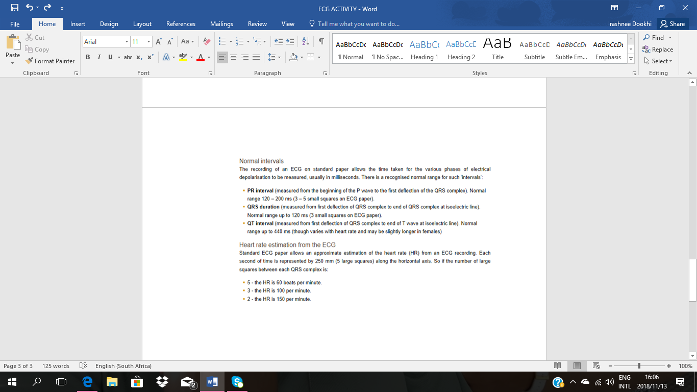
What is the approximate heartrate of the above individual?__________________
MARKING MEMORANDUM
1.1 a) P- ATRIA CONTRACT
b) QRS- VENTRICULAR SYSTOLE
c) T- VENTRICULAR DIASTOLE
d) Q- PRE-VENTRICULAR CONTRACTION
e) R-PEAK OF VENTRICULAR CONTRACTION
f) S- REPOLARISATION OF VENTRICLES
1.2 AFTER SA NODE CONTRACTS
1.3 – MEASURES RHYTHM OF HEARTBEAT
- CAN INDICATE SIZE AND POSITION OF CHAMBERS
- DAMAGE TO MUSCLES
- EFFECT OF DRUGS
- FUNCTIONING OF PACEMAKER
2.1 ELECTRODES
2.2 EACH SHOWS A DIFFERENT REGION OF THE HEART
2.3 CAN INDICATE EXACT AREA THAT IS DAMAGED
2.4 5=60
4,25= x
51








 Бұл курс Қазақстан Республикасы Оқу-ағарту министрлігімен келісілген